Help with a repaired cleft lip/palate patient who is still drooling

Help with a repaired cleft lip/palate patient who is still drooling
I need help with a repaired cleft lip/palate patient who is still drooling. I’ve been working with him for several months – lip strengthening, resting tongue posture, and more. I feel like I’m spinning my wheels trying to get him to habituate lips together. He can do this without much tension…his lower lip, to me is very “droopy” and weak. What can I do? He’ll be 10 years old in October…but is quite immature and struggles with practicing his exercises when I’m not there…per his mother. I’m going to put a list together this week with reminders to help him get the connection from his mouth to his brain. He is a sweetie tho!! I love my patients 😉
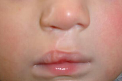
Answer Provided by Dr. Robert M. Mason, DMD, PhD, CCC/SLP Thanks for your questions. What you are inquiring about has traditionally been a part of speech-language pathology, since all cleft palate and craniofacial teams have a speech-language pathologist on the team, so I appreciate that you have not had formal training in this area of therapy. This is certainly not a traditional area that has involved orofacial myofunctional therapy. Nonetheless, what you have been doing appears to be very appropriate to me. Good work! Let’s start first with the “droopy” and weak lower lip. I would describe this situation with your patient as a flaccid, everted lower lip. This is seen in many patients with a repaired bilateral cleft lip, but can also be seen in some patients with a unilateral cleft lip. The reason for this in either case (especially in patients with bilateral clefts) is a lack of adequate muscle union across the upper lip area. This ends up being a tight upper lip. In the bilateral cleft condition, there are three parts that need to be brought together in the initial cleft lip surgery: the two lateral segments (maxillary processes) where the muscle tissue comes from, and the the midline portion of the lip (medial nasal process) that descends down to fuse with the lateral segments in the early embryo. The middle section does not have any muscle tissue in it, so the surgeon has to separate out and take the orbicularis oris muscles from the lateral segments and bring them across and attach them within the middle of the medial nasal process of the upper lip, then cover them with the tissue and mucosa of the medial nasal process. Due to a paucity of tissue, and since a cleft lip and palate by definition is a hypoplastic condition, the surgeon may not be able to establish a good muscle union across the upper lip area; thus, the development of a flaccid, everted lower lip, and a very tight upper lip. Patients with unilateral cleft lips are less apt to have this problem but still, since clefting involves a lack of sufficient tissue (hypoplasia), surgical union of muscle tissue across the front of the face above the vermillion of the upper lip may also fail to be adequate for a good cosmetic result in patients with a unilateral cleft lip. An easy clinical way to determine whether there is an intact muscle union across the upper lip area is to have the patient exaggerate a sustained “ooo” and “eee” (or in phonetics, /u/ and /i/). As the muscles attempt to contract during these simple tasks, you will see a dimpling in the area where a lack of muscle union is involved. If dimpling is found, your therapy cannot be successful since the top half of the oral sphincter is defective. The clinical guideline is that the way to evaluate an everted, weak lower lip in a patient with a repaired cleft lip is to evaluate the competency of muscle union across the upper lip. When the upper half of the oral sphincter is defective, the lower part will become flaccid and everted. What is often done to remedy this situation is for the plastic surgeon do a muscle transposition surgery, taking a wedge or V-shaped full-thickness flap (all the way through from skin to lip mucosa) of the lower lip tissue, which is then raised, and inserted into the surgical area of the upper lip that is opened up to accept this flap. This surgical procedure is called an Abbe flap. (Surgeon Robert Abbe, in 1898, was the first surgeon to successfully perform a cross-lip flap). The blood supply has to be continuous, and the viability of the flap is dependent on the labial blood vessels. The transposed tissue flap, including the vermillion, is inserted into the upper lip and the orbicularis oris muscles are connected. You can see the surgical connection of the upper and lower lips in the drawing “C”. The flap is sutured in layers to reattach the lateral upper lip muscles with the muscle tissue from the lower lip. The lips are then surgically separated in about 10-12 days. This technique can result in creating a philtrum, and can also create a cosmetically acceptable balance between the two sides. Thus, the muscle segment of the orbicularis oris from the lower lip is incorporated into the muscle units of the orbicularis oris of the upper lip. If you determine that there is a lack of muscle tissue continuing across the midface above the upper lip, and if you have tried diligently to strengthen and tone the lower lip, you can confidently discuss the possible need for this secondary lip procedure with the plastic surgeon involved. Any surgeon would know what this means. In patients with unilateral cleft lip, it is sometimes possible to redo the lip repair as a secondary procedure, as more muscle tissue becomes available over the first few years of life. You also asked about chronic drooling. This is not a usual characteristic associated with a cleft lip or palate condition. Such problems are usually handled by the family physician. .The only thing I would recommend is to simply remind your patient to swallow more often. If he concentrates on keeping his lips closed, this also helps. But if this is a chronic problem that concerns the parents, their physician needs to become involved. A medication to dry up salivary flow would be the first line of response. In some cases, the parotid ducts are surgically transposed posterior ward into the pharynx so that the excess saliva flow is swallowed rather than expressed into the mouth. This operation is called the Wilkie procedure. I found a good resource on the internet which is also attached. Be sure to read the overview and then click on the treatment. This should provide you with sufficient information to help you decide whether this situation in your patient needs to be referred out. I hope that these comments have fully answered your questions about this patient. Thanks for asking for help. That is always a sign of an excellent clinician – to know what questions should be asked, and when. Good luck.

