Labial Frenulae – Important Orthodontic Considerations
Published: May 1, 2023
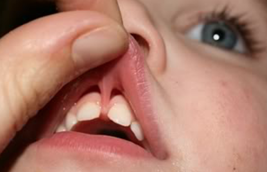
While much appropriate attention has been directed toward the evaluation and treatment of tongue-tie, the labial frenum can also become problematic and may need to be released. Although there are normally several labial-gingival frenulae around the upper and lower labial and buccal vestibules, the midline maxillary and mandibular frenums are the ones that can become problematic.
A short, constricted maxillary midline frenum can interfere with sucking and feeding and later, speech misarticulations and may be indicated for release of the connective tissues involved. A simple “clipping” may be appropriate in early infancy or beyond.
A concern that needs to be addressed, however, is the status of a maxillary or mandibular midline frenum attachment that extends over the alveolar ridge. In the maxillary arch, it is critical to avoid cutting and removing the extra connective tissue at an early age. Such procedures often result in leaving a “window” at the gingival area when the central incisor teeth are fully erupted. The prudent perspective about a maxillary labial frenum that extends over the dental arch is to not remove this extra tissue until the central incisors edges are in contact at the midline. In instances where a diastema develops with dental eruption as a result of the maxillary frenum extending across the alveolus, orthodontic treatment should first be undertaken to close the diastema before surgery to correct the frenum is achieved. Such surgery should involve contouring the connective tissue excess rather than a simple removal of extra tissue. In this way, a “window” at the gum level is avoided, and the contouring provides an acceptable and more ideal cosmetic result.
The mandibular labial midline frenum can also negatively impact sucking, feeding and later speech productions. As well, a frenum that extends across the anterior alveolus can have a negative impact on dental eruption, even causing ectopic eruptions of the central incisors, either lingually or labially. As with a maxillary midline frenum, correction can be accomplished early by clipping the frenum, or releasing it with the laser. Although there is less of a chance that a window will develop following eruption of incisors if the extra frenum tissue is surgically excised, the prudent choice is to wait for eruption of the lower central incisors to undertake a definitive contouring of the area where the connective tissues of the frenum cross the alveolus. However, there seems to be a lesser risk of creating a gingival-located window from surgical removal of the extra frenum tissue. The laser has been used effectively and successfully from infancy onward to contour the gingival area between the central incisors.
For sucking, feeding, and to prevent later speech problems in infancy and childhood, when a short, restricted labial frenum is released by whatever method that is used, the mid-portion of the frenum should be the focus of tissue release. In this way, the tissue mass that extends across the alveolar ridge is preserved for definitive contouring later on following closure of whatever diastema may appear from eruptions of central incisors.
Thank you to Robert M. Mason, DMD, PhD for his article contribution.

